Cells of the body are under the continual forces of compression, tension and shear (collectively, strain.) These mechanical forces play important roles in the regulation of various biological processes such as gene expression, adhesion, migration, and apoptosis. These forces, and the cellular responses, determine the efficacy of medical interventions including pharmaceuticals, tissue implants or external treatments such as physical or laser therapies. The field of Mechanobiology addresses this reaction of cells to applied forces and has been cited in the literature for over 30 years. However, it is just beginning to find its fit in applied healthcare.
To date, there have been limited ways to study the behavior of cells under strain. Academic researchers have developed their own ‘one-off’ devices specific to their experiments. Commercial entities have developed products to measure strain but such devices fail to allow for the measurement of particular forces. These approaches have also been expensive and difficult to adapt to a variety of experimental settings.
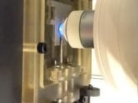
Researchers at Montana State University have developed an inexpensive, versatile and robust device, mountable on a microscope stage, to apply compressive loads to biological samples. Strain is applied via a rotational stepper motor and actuation of the device is controlled by a TTL signal which enables actuation control in conjunction with image acquisition.
Features
• Sterilizable cassette with a disposable cartridge
• Capable of loads up to 150 pounds
• Designed to work with a wide range platforms
Benefits
• In-vitro setting for live cell imaging mimics in-vivo environment
• Inexpensive to manufacture, low cost of ownership to end users
• Wide viewing angle
Uses
• Applying compression to tissue samples or cell seeded constructs – applications in cancer diagnosis, stem cell analysis, cell sorting, osteoarthritis diagnosis
• Live tissue research, development of artificial tissue
• Testing of pharmaceuticals on cells under stress
• Studying the roles of mechanical forces in cellular biology
Technology Transfer and Development Status: A patent is pending and research is ongoing.
This technology comes from the Osteoarthritis and Mechanobiology Lab of Dr. Ron June http://www.mbprogram.montana.edu/faculty.asp?per_id=181&in_id=9
-
Awards
-
 2012 Top 100 Entries
Like this entry?
-
About the Entrant
- Name:Gary Bloomer
- Type of entry:teamTeam members:Ronald June, Ph.D.
Kyle Gunnarson
Jeffrey Runia - Software used for this entry:Solid Works
- Patent status:pending


